To study the biology of species and understand biological diversity, the sampling of wildlife and animal experimentation for research purposes are associated with important ethical and legal questions. One alternative in research is the use of viable animal cells.
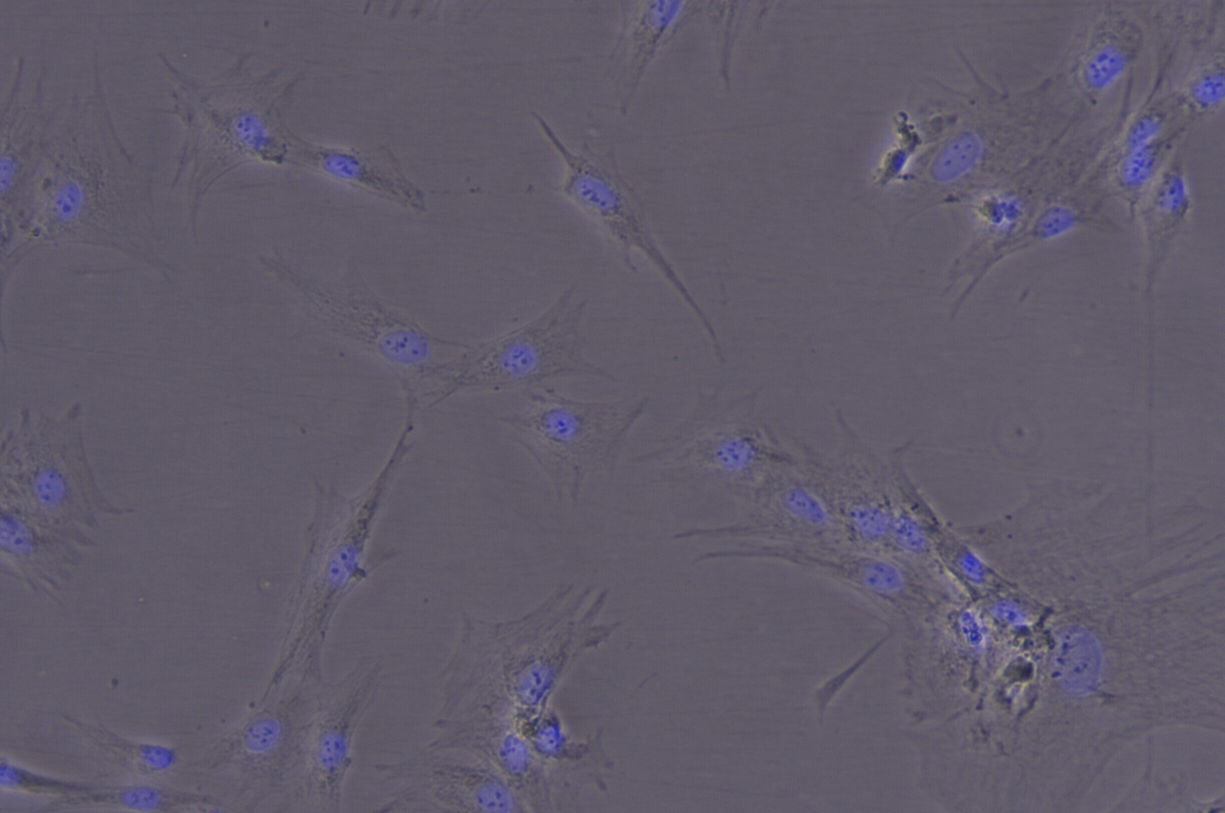
Cell culture from skin tissue of Nile goose (Alopochen aegyptiaca).
Cell culture is one of the most important techniques in biological and medical research. Each individual cell carries identical copies of the genetic information, DNA, which provides crucial molecular information about the respective animal species. It allows the study of metabolism, cell division, and various other cellular processes.
A single unique cell can multiply practically indefinitely while preserving the original genetic information. In other words, an extinct species can continue to exist indefinitely through its frozen cells stored in liquid nitrogen (-196°C), which can be used for future research purposes.
In the FOGS project, we follow this approach to obtain viable cells from rare and endangered animal species. We preserve these cells indefinitely to create a repository of rare genetic and molecular information.
In addition to producing cells for the research community, we aim to utilize the high-quality genetic information obtained from these cells to design precise SNPSTR markers that require high-quality DNA. These markers will be used in species identification efforts to combat illegal trade.
The contact person for cell cultures at the LIB is Dr. Camilla Bruno Di Nizo.
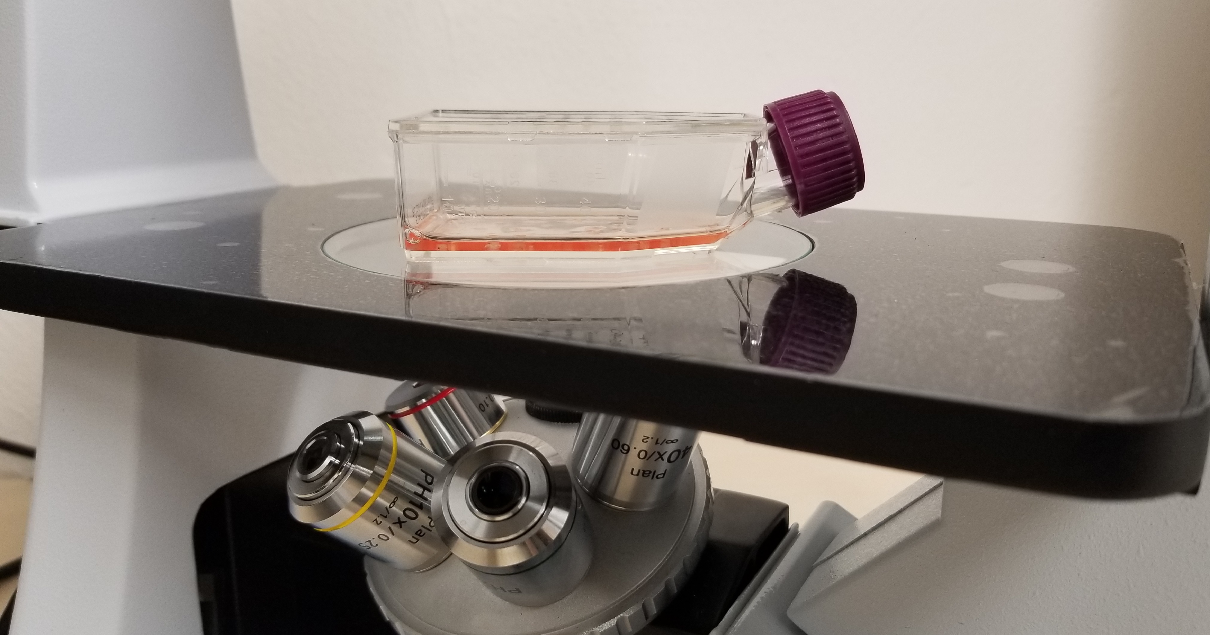
Cell culture flask with culture medium and growing animal cells from tissue pieces. Cell culture laboratory at the ZFMK.
Cell Culture Images
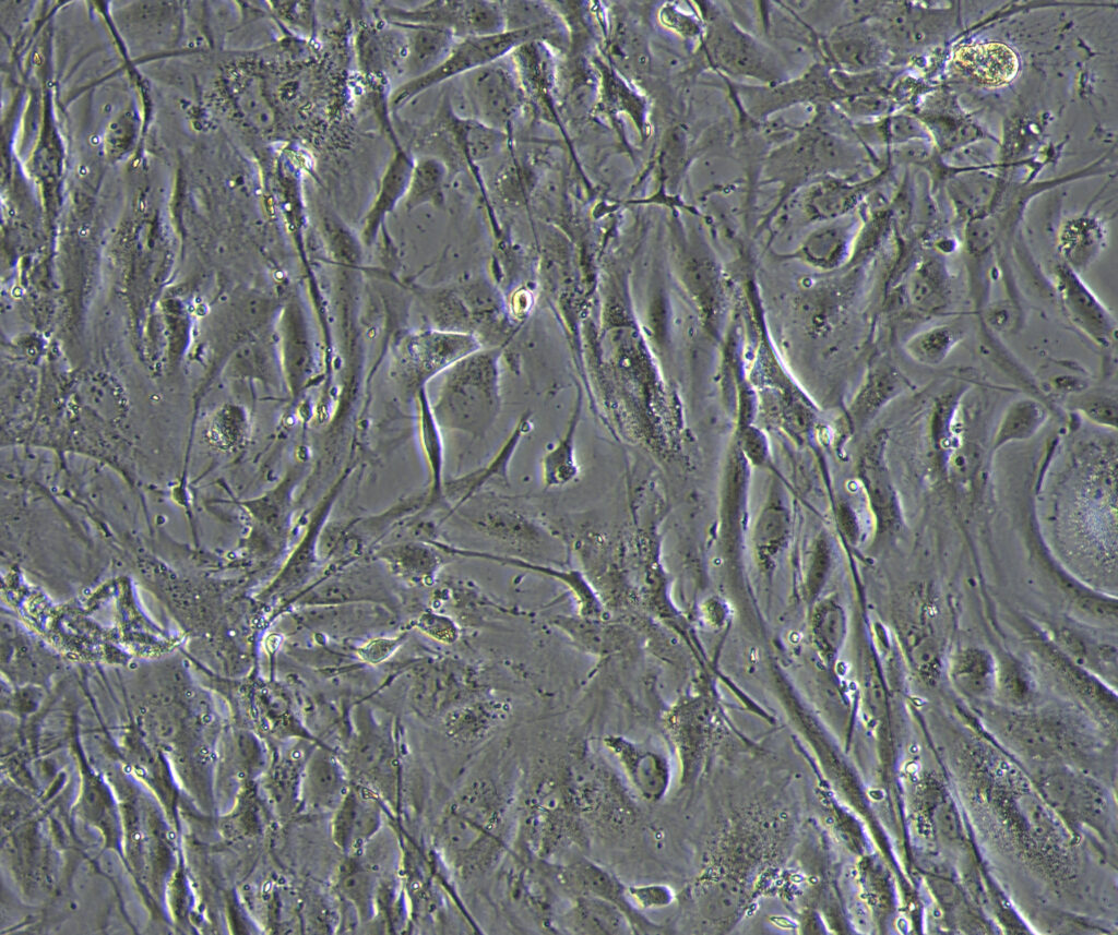
Cell population derived from leg pieces of Bombina variegata (Yellow-bellied toad). One could notice a mix of morphologically different cells that are, inter-alia, muscular syncytium, fibroblast and epithelial-like cells.
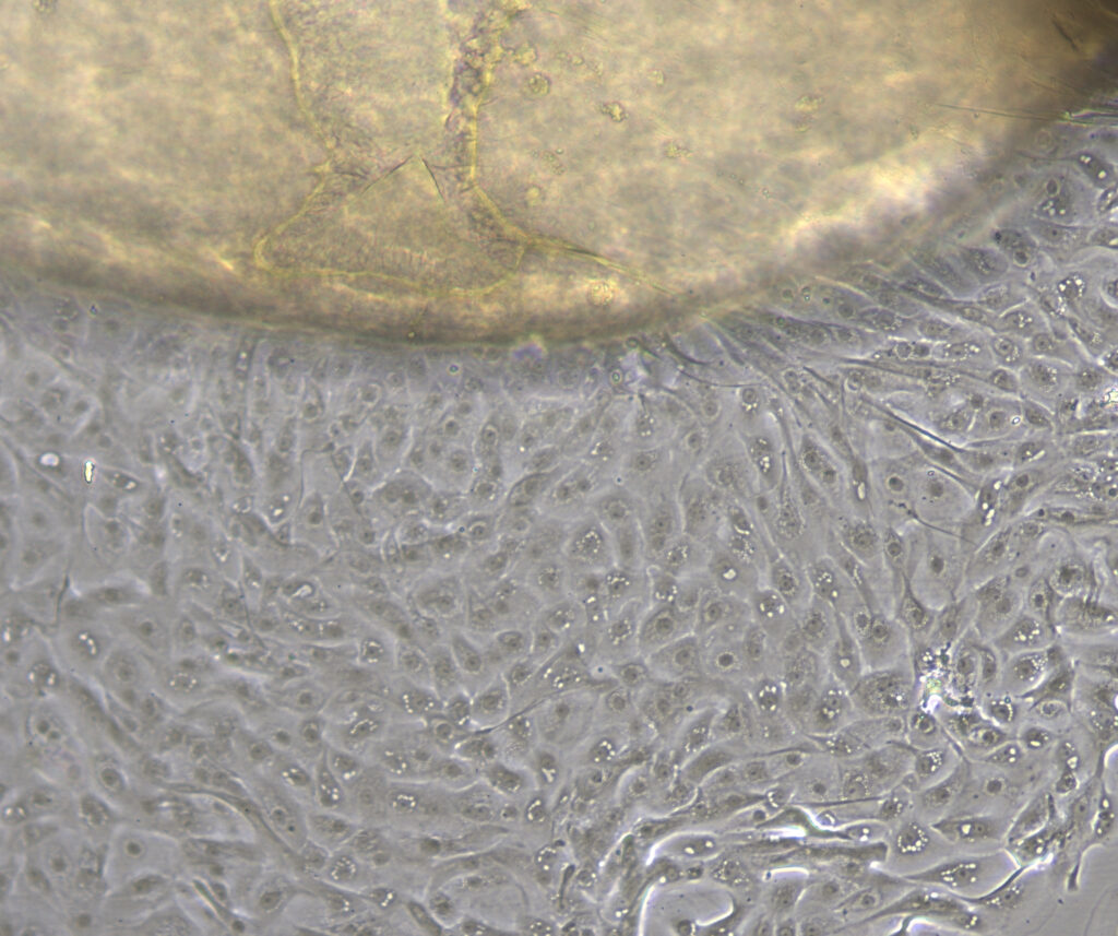
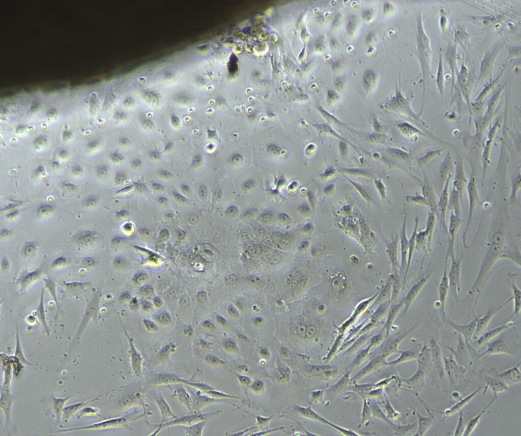
Cell layer growing from an explanted Cornea (structure on the top in the image) collected from a freshly deceased juvenile Falco tinnunculus (Common kestrel). The uniform cell population was obtained after 5 days in culture.
Heterogeneous cell population growing from an explant of lip (structure on the top in the image) collected from a Panthera tigris (Tiger). Epithelial-like cells are growing close to the tissue, while fibroblast-like cells are migrating away in the outer layer.